The Structure and Function of the Human Heart
The human heart is a pump. It pumps blood around the body at different speeds and at different pressures according go the body's needs. It can do this because the wall of the heart is made from cardiac muscle.
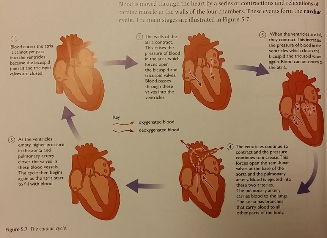
Blood is moved through the heart by a series of contractions and relaxations of cardiac muscle in the walls of the four chambers. These events form the cardiac cycle.
The Cardiac Cycle
When a chamber of the heart is contracting, we say it is in systole.
When it is relaxing, we say it is in diastole.
When it is relaxing, we say it is in diastole.
The Structure of the Heart is adapted to its function in several ways:
- It is divided into a left side and a right side by the septum. The right ventricle pumps blood only to the lungs while the left ventricle pumps blood to all other parts of the body. This requires much more pressure, which is thy the wall of the left ventricle is much thicker than that of the right ventricle.
- Valves ensure that blood can flow only in one direction through the heart.
- The walls of the atria are thin. They can be stretched to receive blood as it returns to the heart but can contract with enough force to push blood through the bicuspid and tricuspid valves into the ventricles.
- The walls of the heart are made of cardiac muscle which can contract and then relax continuously, without becoming fatigued.
- The cardiac muscle has its own blood supply called the coronary circulation.
- Blood reaches the muscle via coronoary arteries. These carry blood to cappilaries that supply the heart muscle with oxygen and nutrients. Blood is returned to the right atrium via coronary veins.
Heart Rate
The heart beats about 70 times a minute, but this can change according to circumstances.
When we exercise, muscles must release more energy. THey need and increased supply of oxygen for aerobic respiration. TO deliver the extra oxygen, both the number of beats per minute (heart rate), and the volume of blood pumped with each beat (stroke volume) increase.
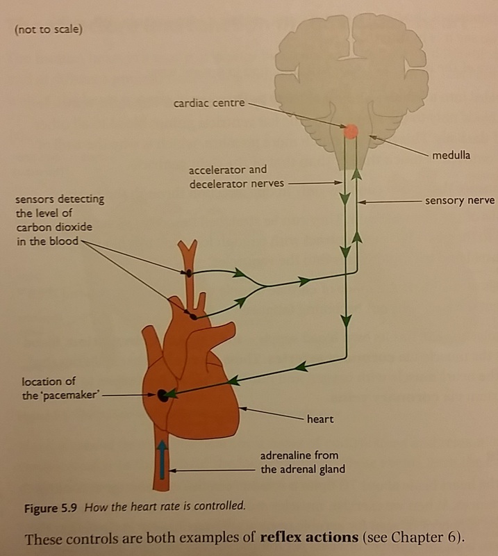
These changes in the heart rate are brought about by nerve impulses from a part of the brain called the medulla. When we start to exercise, our muscles produce more carbon dioxide in aerobic respiration. Sensors in the aorta and the carotid artery (the artery leading to the head) detect this increase. They send nerve impulses to the medulla. The medulla responds by sending nerve impulses along the accelerator nerve. When carbon dioxide production returns to normal, the medulla receives fewer impulses. It responds by sending nerve impulses along a decelerator nerve.
The accelerator nerve increases the heart rate. It also causes the heart to beat with more force and so increases blood pressure.
The decelerator nerve decreases the heart rate. It also reduces the force of the contractions. Blood pressure then returns to normal.
When we exercise, muscles must release more energy. THey need and increased supply of oxygen for aerobic respiration. TO deliver the extra oxygen, both the number of beats per minute (heart rate), and the volume of blood pumped with each beat (stroke volume) increase.
These changes in the heart rate are brought about by nerve impulses from a part of the brain called the medulla. When we start to exercise, our muscles produce more carbon dioxide in aerobic respiration. Sensors in the aorta and the carotid artery (the artery leading to the head) detect this increase. They send nerve impulses to the medulla. The medulla responds by sending nerve impulses along the accelerator nerve. When carbon dioxide production returns to normal, the medulla receives fewer impulses. It responds by sending nerve impulses along a decelerator nerve.
The accelerator nerve increases the heart rate. It also causes the heart to beat with more force and so increases blood pressure.
The decelerator nerve decreases the heart rate. It also reduces the force of the contractions. Blood pressure then returns to normal.
The precise region of the medulla that controls heart functions is called the cardiac centre.